


Biblioteka Théa

Unikalna kolekcja publikacji poświęconych różnej tematyce i schorzeniom okulistycznym, opracowanych we współpracy z uznanymi międzynarodowymi ekspertami w tej dziedzinie.

Witamy w Bibliotece Théa – bibliotece okulistycznej, oferującej wyjątkową cyfrową kolekcję publikacji autorstwa wybitnych międzynarodowych ekspertów.

Biblioteka Théa to cyfrowa kolekcja  medyczna starannie opracowana przez firmę Théa, której celem jest wspieranie profesjonalnej edukacji w dziedzinie okulistyki. Zawiera wysokiej jakości, bogato ilustrowane książki oraz przewodniki kliniczne, przygotowane we współpracy z renomowanymi okulistami z całego świata.
Tematyka obejmuje kluczowe obszary, takie jak: OCT i jaskra, przewlekłe owrzodzenia rogówki, guzy powiek i spojówek a także historyczny rozwój narzędzi i metod leczenia okulistycznego.

Dostępna poprzez platformę Théa Academy biblioteka, wspiera specjalistów zajmujących się opieką okulistyczną — od lekarzy w trakcie specjalizacji po doświadczonych praktyków — w zdobywaniu aktualnej wiedzy, poznawaniu najnowszych osiągnięć klinicznych i odkrywaniu historycznych aspektów okulistyki.

Loading…
Nowość!

What’s up with your eye?

Pr. Marie-José TASSIGNON / Pr. Nicoline SCHALIJ-DELFOS,
Inne
Early vision screening is essential for children’s eye health and overall development. Eye conditions such as myopia or amblyopia (lazy eye) can go unnoticed, but if left undetected, they may negatively impact school performance, self-confidence, and quality of life. Systematic eye screening in schools and community settings enables early detection and timely treatment, which is essential to prevent permanent vision impairment and promote healthy development.
Zobacz więcej
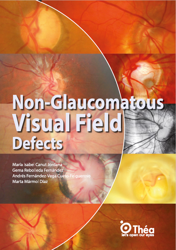
Non glaucomatous visual field defects

Maria Isabel CANUT JORDANA / Gema REBOLLEDA FERNANDEZ / Andrés FERNANDEZ-VEGA CUETO-FELGUEROSO / Marta MARMOL DIAZ,
Jaskra
Non-glaucomatous visual field defects are alterations in visual perception not caused by glaucomatous damage to the optic nerve. This book highlights the importance of distinguishing these from typical glaucomatous field loss, as they often stem from neuro-ophthalmological or congenital conditions, each requiring distinct diagnostic and therapeutic strategies. Common conditions producing these defects include optic disc drusen, tilted disc syndrome, optic nerve hypoplasia, optic disc coloboma, and morning glory disc anomaly.  
Zobacz więcej

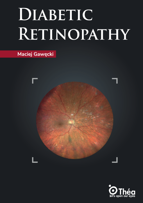
Diabetic Retinopathy

Maciej GAWECKI,
Siatkówka
Diabetic retinopathy is one of the most serious complications of diabetes and a leading diabetes-related eye disease, affecting the retina, the delicate tissue at the back of the eye that is essential for vision. High blood sugar levels over time cause progressive retinal damage by harming the small blood vessels in the retina, leading them to leak fluid or bleed. This process can result in swelling (macular edema), the formation of abnormal new blood vessels (proliferative diabetic retinopathy), and, eventually, progressive vision impairment.
Zobacz więcej

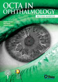
OCTA in ophthalmology

Angela CARNEIRO / Rita FLORES / Antonio CAMPOS,
Obrazowanie Okulistyczne
Optical Coherence Tomography Angiography (OCTA) is a cutting-edge, non-invasive method that enables detailed visualization of the retinal and choroidal microvasculature without the need for dye injections. OCTA in ophthalmology has emerged as a revolutionary imaging technique, providing depth-resolved, high-resolution imaging of retinal blood flow by detecting motion contrast from red blood cells. Unlike traditional fluorescein angiography (FA), which is limited to two-dimensional imaging and is affected by leakage and staining, optical coherence tomography angiography allows clinicians to assess both the structure and function of the eye’s microvascular networks in real time.
Zobacz więcej

Glaucoma stories

Florent APTEL / Jean-François ROULAN,
Jaskra
This glaucoma review provides insights into various presentations of the disease through 20 real-world clinical cases. The most common form primary open angle glaucoma is a recurrent theme, though rarer forms such as angle closure glaucoma and secondary glaucomas (e.g., uveitic, pigmentary, steroid-induced) are richly illustrated. A unifying thread is the progressive and irreversible nature of visual field defects, reinforcing the urgency of early diagnosis. Several cases show how ocular hypertension, initially detected incidentally, evolves silently before clinical symptoms emerge.
Zobacz więcej

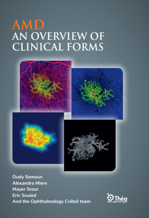
AMD an overview of clinical forms

Oudy SEMOUN / Alexandra MIERE / Mayer SROUR / Eric SOUIED,
Siatkówka
Age-related macular degeneration (AMD) is a leading cause of vision loss in the elderly, affecting the central portion of the retina and progressively impairing central vision. This retinal disease manifests in two primary AMD clinical forms: dry (atrophic) and wet (exudative). Dry AMD, also known as atrophic AMD, involves gradual retinal pigment epithelium degeneration and the accumulation of drusen yellow deposits beneath the retina. This form accounts for approximately 85–90% of AMD cases. It typically progresses slowly and can culminate in geographic atrophy, leading to central vision loss.  
Zobacz więcej

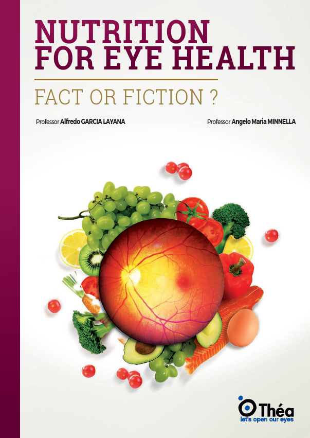
Nutrition for eye health

Alfredo GARCIA LAYANA / Angelo MARIA MINNELLA ,
Suplementacja
The role of nutrition in maintaining ocular health is increasingly recognized as crucial for both preserving vision and preventing eye diseases. The eye, as a highly metabolically active organ, requires a constant supply of specific nutrients to maintain its structure and function. There is evidence suggesting that essential nutrients such as vitamins A, C, and E, zinc, omega-3 fatty acids, lutein, and zeaxanthin may assist in protecting ocular tissues and supporting their natural regenerative processes. 
Zobacz więcej

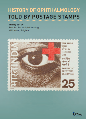
History of ophthalmology told by postage stamps

Thierry ZEYEN,
Inne
The anatomical conception of the eye has undergone a remarkable transformation throughout history. Early records, such as the Ebers Papyrus from 1550 B.C., reveal how ancient civilizations blended myth, medicine, and philosophy in their understanding of anatomy and physiology. In those times, the eye was often regarded as a mystical organ, with healing powers attributed to symbols like the Eye of Horus, a motif prominently featured on stamps from the 1937 Cairo Congress on ophthalmology. This symbol not only represented protection and health but also reflected the deep cultural significance of vision in ancient societies.
Zobacz więcej

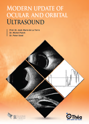
Modern update of ocular and orbital ultrasound

Mario DE LA TORRE / Peter GOOD / Michel PUECH,
Obrazowanie Okulistyczne
Ocular and orbital ultrasound is a non-invasive imaging technique that utilizes high frequency sound waves to visualize the structures of the eye and surrounding orbital tissues. This method is particularly valuable when direct examination is hindered by media opacities, such as dense cataracts, vitreous hemorrhages, or corneal scars.
Zobacz więcej

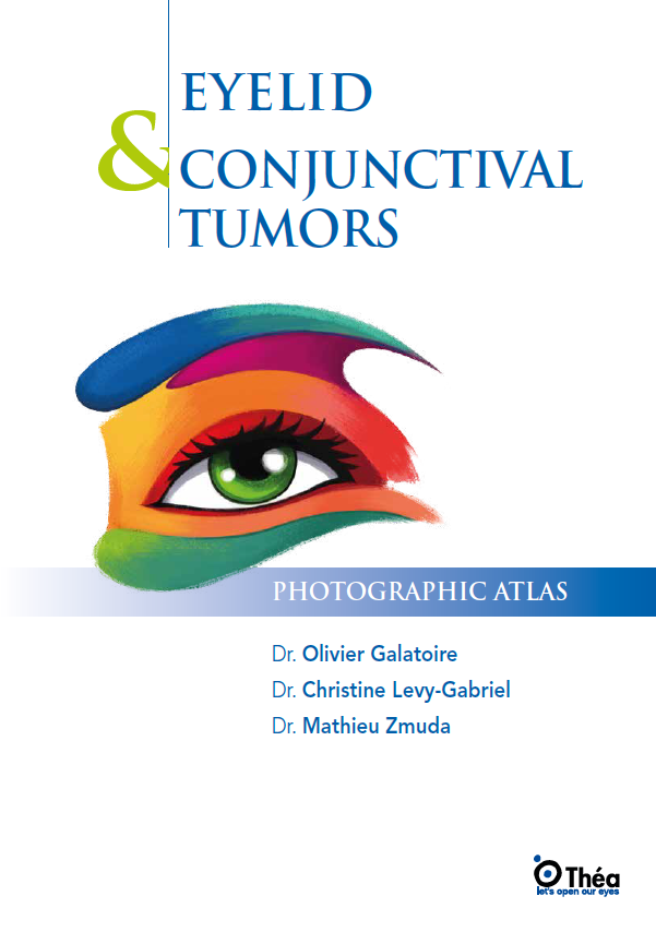
Eyelid and conjunctival tumors: a comprehensive guide for ophthalmologists

Olivier GALATOIRE / Christine LEVY-GABRIEL / Mathieu ZMUDA,
Powieki
The eyelid and conjunctiva are critical components of the ocular system, performing essential functions such as protecting the eye, regulating corneal hydration, and contributing to eye aesthetics.
Zobacz więcej

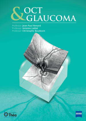
OCT and glaucoma: leveraging imaging for early diagnosis and management

Jean-Paul RENARD / Antoine LABBÉ / Christophe BAUDOUIN,
JaskraObrazowanie Okulistyczne
Optical Coherence Tomography (OCT) has revolutionized the diagnosis and management of glaucoma, providing clinicians with precise and reproducible imaging of ocular structures. OCT uses light waves to take cross-section pictures of the retina and optic nerve head (ONH). It has become an indispensable tool in the early detection of glaucomatous optic neuropathy, enabling the identification of structural damage before significant visual field defects manifest.
Zobacz więcej

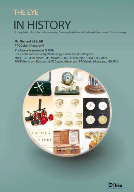
The eye in history: a journey through instruments, diagnostics, and clinical progress

Richard KEELER / Harminder S DUA,
Inne
The evolution of ophthalmology is deeply intertwined with the development of various instruments that have revolutionized the practice. From the early days of the ophthalmoscope, invented by Hermann von Helmholtz in 1850, to the intricate devices used today, each instrument has played a critical role in enhancing our understanding and treatment of the eye. The direct ophthalmoscope, created by Helmholtz, allowed for the first detailed examination of the interior of the eye, laying the groundwork for modern ophthalmology. The subsequent development of the binocular indirect ophthalmoscope by Marc-Antoine Giraud-Teulon and others further expanded the capabilities of eye examination, enabling the visualization of the fundus in three dimensions. These advancements illustrate the shift from theoretical concepts to practical applications in eye history.
Zobacz więcej
PRZETŁUMACZ »